•Anatomically the cortex is divided into 6 lobes: frontal, parietal, temporal, occipital, limbic and insular
•Each lobe has several gyri
•Functionally the cortex is divided into numbered areas first proposed by Brodmann in 1909
•Brodmann’s areas were described based on cytoarchitecture (density and numbers of different cortical neurons and thickness ); later they were found to be functionally significant
Functional Areas of the Cerebral Cortex
•The three types of functional areas are:
–Motor areas – control voluntary movement
–Sensory areas – conscious awareness of sensation
–Association areas – integrate diverse information
–Most of the human brain cortex is association cortex
•Each hemisphere is chiefly concerned with the sensory and motor functions of the opposite (contralateral) side of the body
•Symmetrical in structure but two hemispheres are NOT entirely equal in function
Cerebral Cortex: Motor Areas
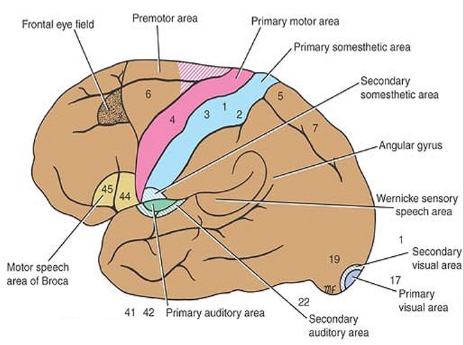
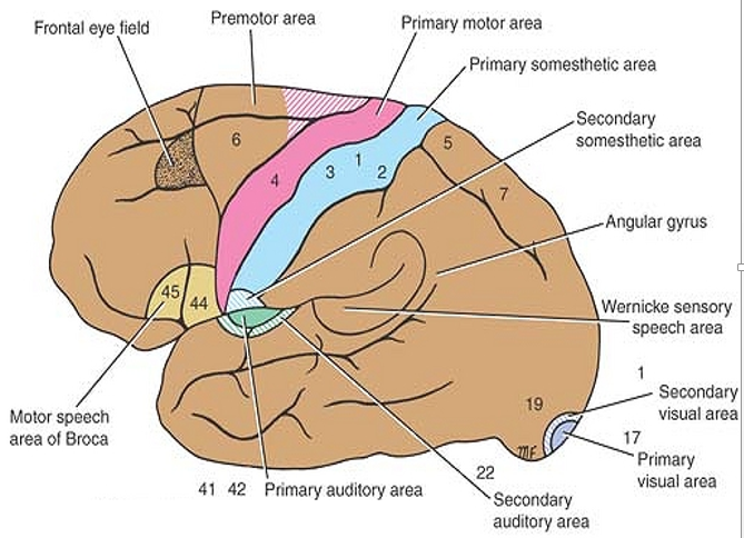
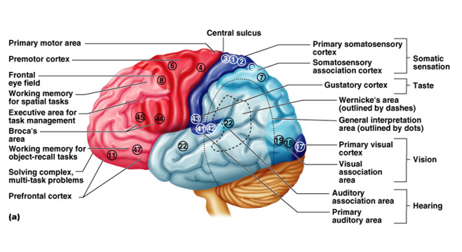
•Primary motor area ( area 4)
•Premotor ( secondary ) motor area ( area 6)
•Supplementary motor cortex (parts of areas 6 & 8)
•Frontal eye field (area 8 )
•Broca’s area (44 & 45)
Primary Motor Cortex
•Located in the precentral gyrus & anterior part of paracentral lobule
•Composed of pyramidal cells whose axons make up the corticospinal tracts
•Its function is to carry out the individual movements of different parts of the body
•Allows conscious control of precise, skilled, voluntary movements
•Motor innervation is contralateral
•The movement areas of the body are represented in inverted form in the precentral gyrus
Premotor ( secondary ) motor area
•Located just anterior to the precentral gyrus
•Controls learned, repetitious, or patterned motor skills (typing, playing musical instrument)
•Coordinates sequential actions
•Involved in the planning of movements
Supplementary motor cortex,
– parts of areas 6 & 8, programming for
complex movements
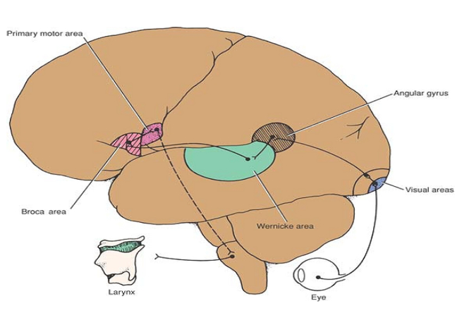
Broca’s Area
•Broca’s area
–Located anterior to the inferior region of the premotor area( inferior frontal gyrus)
–Present in one hemisphere (usually the left)
–A motor speech area that directs muscles of the tongue
–Is active as one prepares to speak
–If lost in early childhood (trauma, tumors) may develop in right hemisphere
–Dysfunction leads to aphasia (expressive aphasia)
Frontal eye field, area 8:
Saccade is a quick movement of both eyes between two or more phases of fixation , In contrast, in smooth pursuit movements the eyes move smoothly instead of in jumps
– The FEF is responsible for saccades for the purpose of visual field perception and awareness, its independent of visual stimuli
Cerebral Cortex: Sensory Areas
•Primary somatosensory area
•Somatosensory association area
•Visual and auditory areas
•Olfactory, gustatory, and vestibular areas
Primary Somatosensory Area
•Located in the postcentral gyrus posterior part of the paracentral lobule
• this area:
–Receives sensory information from the skin and skeletal muscles via dorsal column and spinothalamic tracts
–able to identify body region being stimulated
• The opposite half of the body is represented as inverted. the size of the cortical area is proportional to the number of sensory receptors present in that part of the body
Somesthetic association area , areas 5, 7:
•Its function is to receive and integrate different sensory modalities.
•Inputs in these areas allows for the perception of shape, size, and texture and the identification of objects by contact (stereognosis)
•May assist with visuo-motor coordination.
•Bilateral lesions in the somesthetic association
areas in humans are associated with inability to move the hand
toward an object that is clearly seen
– Unilateral lesions in (non-the hemisphere) produce neglect of the contralateral half of the body and visual space.
Functional Areas of the Cerebral Cortex

Secondary somatosensory cortex:
Inferior aspect of postcentral gyrus and in the superior bank and depth of the lateral sulcus
-Body representation in this area is bilateral, with contralateral predominance
-The secondary somesthetic area contains no cells sensitive to joint movement or joint position
– Its function is not clear
Gustatory area
-In the most inferior part of the postcentral gyrus of the parietal lobe (insula , and opercula of frontal )
-Primary site involved with the interpretation of the sensation of Taste.
•Primary auditory area
•Located at the superior margin of the temporal lobe
–Receives information related to pitch, rhythm & loudness of sound
•Auditory association area
–Located posterior to the primary auditory cortex
–Stores memories of sounds and permits perception of sounds
. Primary olfactory (smell) area
– In the piriform and periamygdaloid regions of the temporal lobe
– Interprets the sense of smell.
• vestibular area is believed to be situated near the part of the postcentral gyrus concerned with sensations of the face.
Parietal Neglect Syndrome Clinical Illustration

- •Failure to recognize side of body contralateral to injury
- •May not bathe contralateral side of body or shave contralateral side of face
sensory speech area of Wernicke
•is localized in the left dominant hemisphere, mainly in the superior temporal gyrus, with extensions around the posterior end of the lateral sulcus into the parietal region. The Wernicke area permits the understanding of the written and spoken language and enables a person to read a sentence, understand it, and say it out loud
Aphasia
A defect in language processing caused by dysfunction of the dominant
cerebral hemisphere.
Broca’s Aphasia ( motor)
Wernicke’s Aphasia
Global aphasia ( both)
Angular gyrus ( part of sensory area of speach:
Damge lead to unable to read ( alexia) & to write ( agraphia)
Primary visual cortex, upper and lower banks of calcarine fissure, area 17
Receives visual information from the retinas
Secondary visual cortex, areas 18, 19; storage past visual experiences, thus enabling the individual to recognize and appreciate what he or she is seeing.
Posterior eyes field :
The occipital eye fields are not as well defined as the frontal eye field. T
– located in the region near the junction of the occipital lobe with the posterior parietal and temporal lobes
– Its function is reflexive movements of the eye when it is following an object

the output from the primary visual cortex follows two path-ways :
to the occipitoparietal cortex(the “where” pathway) and to the occipitotemporal cortex (the “what” pathway)
Bilateral lesions in the “where” pathway result in the inability to direct the eyes to a certain point in the visual field with intact eye movements
Bilateral lesions in the “what” pathway result in the
inability of patients, with normal visual perception, to compre-
hend the meaning of nonverbal visual stimuli (visual agnosia)
Association Areas
•Cortical areas of the cerebrum with complex functions
•Include:
–Prefrontal cortex
–Language areas
–General (common) interpretation area
–Visceral association area
Association Areas
I. Prefrontal Cortex
•Located in the anterior portion of the frontal lobe
•Involved with intellect, cognition, recall, and personality
•Necessary for judgment, reasoning, persistence, and conscience;
–These qualities develop slowly in children implying that the prefrontal cortex matures slowly; heavily dependent on positive and negative feedback from one’s social environment
•Closely linked to the limbic system (emotional part of the brain) Tumors/lesions cause mental and personality disorders: mood swings, loss of judgment, attentiveness, and inhibitions
Association Areas
II. Language Areas
•Located in a large area surrounding the left (or language-dominant) lateral sulcus
•Major parts and functions:
–Wernicke’s area – involved in sounding out unfamiliar words
–Broca’s area – speech preparation and production
•Aphasias: defects in language
•Broca’s area: expressive aphasia; have trouble forming words and putting them in grammatical order
•Wernike’s area: comprehensive aphasia; difficulty understanding spoken /written language;
Association Areas
III. General Interpretation Area
•Ill-defined region; parts of temporal, parietal, and occipital lobes
•In one hemisphere only
•Receives input from all sensory association areas and integrates them into a single thought or understanding of the situation
E.g., dropping a bottle of acid in lab “danger”
Association Areas
IV. Visceral Association Area
•Cortex of the insula
• May be involved in conscious perception of visceral sensations
dominant hemisphere. The left half of the brain in almost all right-handed people and 85% of left-handed people. This is the hemisphere concerned with language , logical thought ,mathematical abilities & areas of behavior.
The non-dominant hemisphere is responsible for creativity, including art , imagination, recognition of faces & music interpretation.